ARP1000-64
[Polyclonal Antibody]
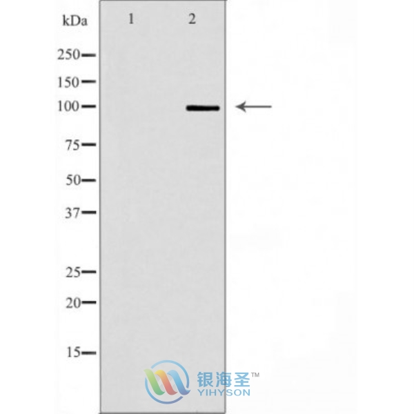
CDH2 Rabbit Polyclonal Antibody
www.yhsbio.com
market@yhsbio.com
support@yhsbio.com
+86-21-54651191
Room 703,Building 6,333# Guiping
Rd.,Xuhui District,Shanghai,China
market@yhsbio.com
support@yhsbio.com
+86-21-54651191
Room 703,Building 6,333# Guiping
Rd.,Xuhui District,Shanghai,China
DATASHEET
| Species: | Rabbit |
| Applications: | WB |
| Immunogen Range: | A synthetic peptide of human CDH2 |
| Clonality: | Polyclonal Antibody |
| Isotype: | IgG |
| GENE ID: | 1000 |
| Swiss Prot: | P19022 |
| Synonyms: | CDHN, NCAD, CD325, CDw325, N-Cadherin |
| Purification: | Affinity purification |
| Storage: | Store at -20°C or -80°C in PBS with 0.02% sodium azide and 5388% glycerol. Avoid freeze/thaw cycles. |
| Background: | Cadherins are a superfamily of transmembrane glycoproteins that contain cadherin repeats of approximately 100 residues in their extracellular domain. Cadherins mediate calcium-dependent cell-cell adhesion and play critical roles in normal tissue development (1). The classic cadherin subfamily includes N-, P-, R-, B-, and E-cadherins, as well as about ten other members that are found in adherens junctions, a cellular structure near the apical surface of polarized epithelial cells. The cytoplasmic domain of classical cadherins interacts with β-catenin, γ-catenin (also called plakoglobin), and p120 catenin. β-catenin and γ-catenin associate with α-catenin, which links the cadherin-catenin complex to the actin cytoskeleton (1,2). While β- and γ-catenin play structural roles in the junctional complex, p120 regulates cadherin adhesive activity and trafficking (1-4). Investigators consider E-cadherin an active suppressor of invasion and growth of many epithelial cancers (1-3). Recent studies indicate that cancer cells have up-regulated N-cadherin in addition to loss of E-cadherin. This change in cadherin expression is called the "cadherin switch". N-cadherin cooperates with the FGF receptor, leading to overexpression of MMP-9 and cellular invasion (3). Research studies have shown that in endothelial cells, VE-cadherin signaling, expression, and localization correlate with vascular permeability and tumor angiogenesis (5,6). Investigators have also demonstrated that expression of P-cadherin, which is normally present in epithelial cells, is also altered in ovarian and other human cancers (7,8). |
| Caculated MW: | 100 kDa |
| Observed MW: | Refer to Figures |
| Applications: |
WB 1:500-1:1000 |
| Reacitivity: | Human, Mouse, Rat |
For research use only. Not intended for diagnostic or therapeutic use!
Additional information