ARP3662-62
[Polyclonal Antibody]
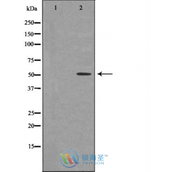
IRF4 Rabbit Polyclonal Antibody
www.yhsbio.com
market@yhsbio.com
support@yhsbio.com
+86-21-54651191
Room 703,Building 6,333# Guiping
Rd.,Xuhui District,Shanghai,China
market@yhsbio.com
support@yhsbio.com
+86-21-54651191
Room 703,Building 6,333# Guiping
Rd.,Xuhui District,Shanghai,China
DATASHEET
| Species: | Rabbit |
| Applications: | WB IHC |
| Immunogen Range: | A recombinant protein of human IRF4 |
| Clonality: | Polyclonal Antibody |
| Isotype: | IgG |
| GENE ID: | 3662 |
| Swiss Prot: | Q15306 |
| Synonyms: | IRF4,LSIRF,MUM1 |
| Purification: | Affinity purification |
| Storage: | Store at -20°C or -80°C in PBS with 0.02% sodium azide and 50% glycerol. Avoid freeze/thaw cycles. |
| Background: | Interferon regulatory factors (IRFs) comprise a family of transcription factors that function within the Jak/Stat pathway to regulate interferon (IFN) and IFN-inducible gene expression in response to viral infection (1). IRFs play an important role in pathogen defense, autoimmunity, lymphocyte development, cell growth, and susceptibility to transformation. The IRF family includes nine members: IRF-1, IRF-2, ISGF3γ/p48, IRF-3, IRF-4 (Pip/LSIRF/ICSAT), IRF-5, IRF-6, IRF-7, and IRF-8/ICSBP. All IRF proteins share homology in their amino-terminal DNA-binding domains. IRF family members regulate transcription through interactions with proteins that share similar DNA-binding motifs, such as IFN-stimulated response elements (ISRE), IFN consensus sequences (ICS), and IFN regulatory elements (IRF-E) (2). IRF-4 was independently cloned by three groups and demonstrated to have roles in different contexts of lymphoid regulation (3-5). First, IRF-4 (Pip) was found to associate with PU.1, a hematopoietic specific member of the ETS family, and to regulate the expression of B-cell specific genes (3). Second, it was characterized as a lymphoid-specific member of the IRF family (LSIRF) and able to bind to ISRE (4). Third, it was identified in activated T cells as a factor that binds to the promoter of the interleukin-5 gene (ICSAT), and shown to repress gene activation induced by IFN (5). IRF-4 is expressed in all stages of B cell development and in mature T cells, and is inducible in primary lymphocytes by antigen mimetic stimuli such as Concavalin A, CD3 crosslinking, anti-IgM and PMA treatment (4,5). Mice deficient in IRF-4 show normal distribution of B and T lymphocytes at 4 to 5 weeks, but later develop progressive generalized lymphadenopathy, suggesting a role for IRF-4 in the function and homeostasis of mature B- and T-lymphocytes (6). |
| Caculated MW: | 52 kDa |
| Observed MW: | Refer to Figures |
| Applications: |
WB 1:500-1:2000 IHC 1:50-1:200 |
| Reacitivity: | Human, Mouse, Rat |
For research use only. Not intended for diagnostic or therapeutic use!
Additional information