ARP7132-01
[Polyclonal Antibody]
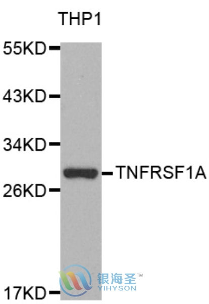
TNFRSF1A Rabbit Polyclonal Antibody
www.yhsbio.com
market@yhsbio.com
support@yhsbio.com
+86-21-54651191
Room 703,Building 6,333# Guiping
Rd.,Xuhui District,Shanghai,China
market@yhsbio.com
support@yhsbio.com
+86-21-54651191
Room 703,Building 6,333# Guiping
Rd.,Xuhui District,Shanghai,China
DATASHEET
| Species: | Rabbit |
| Applications: | WB IHC IF |
| Immunogen Range: | A recombinant protein of human TNF-R1 |
| Clonality: | Polyclonal Antibody |
| Isotype: | IgG |
| GENE ID: | 7132 |
| Swiss Prot: | P19438 |
| Synonyms: | TNF-R1, TNFRSF1A, TNFR1, CD120a, TNF R55, FPF |
| Purification: | Affinity purification |
| Storage: | Store at -20°C or -80°C in PBS with 0.02% sodium azide and 50% glycerol. Avoid freeze/thaw cycles. |
| Background: | TNF-α is an important cytokine produced by numerous cell types including neutrophils, activated lymphoctyes, macrophages an NK cells. It plays a critical role in inflammatory responses and in apoptosis (1). TNF-α exists as a membrane-anchored and soluble form, both of which show biological activity. Response to TNF-α is mediated through two receptors, TNF-R1, which is widely expressed, and TNF-R2, which is expressed mainly in immune and endothelial cells (2). Antagonists to TNF-α have been validated as therapeutic targets for rheumatoid arthritis and other immune disorders (3). The two receptors for TNF-α, TNF-R1 (55 kDa) and TNF-R2 (75 kDa) can mediate distinct cellular responses (4,5). In most cases cytotoxicity elicited by TNF has been reported to act through TNF-R1 (6,7). Cytotoxicity is mediated by a "death domain" with the intracellular region of the receptor that binds to the death domain adaptor protein TRADD and triggers the activation of caspases (8). Soluble forms of both receptors have also been characterized which can bind TNF-α and may play an important role in immune disorders (9,10). |
| Caculated MW: | 50 kDa |
| Observed MW: | Refer to Figures |
| Applications: |
WB 1:500-1:2000 IHC 1:50-1:200 IF 1:50-1:200 |
| Reacitivity: | Human, Mouse, Rat |
For research use only. Not intended for diagnostic or therapeutic use!
Additional information