ARP4772-01
[Polyclonal Antibody]
NFATC1 Rabbit Polyclonal Antibody
www.yhsbio.com
market@yhsbio.com
support@yhsbio.com
+86-21-54651191
Room 703,Building 6,333# Guiping
Rd.,Xuhui District,Shanghai,China
market@yhsbio.com
support@yhsbio.com
+86-21-54651191
Room 703,Building 6,333# Guiping
Rd.,Xuhui District,Shanghai,China
DATASHEET
| Species: | Rabbit |
| Applications: | WB IHC IF |
| Immunogen Range: | A recombinant protein of human NFATC1 |
| Clonality: | Polyclonal Antibody |
| Isotype: | IgG |
| GENE ID: | 4772 |
| Swiss Prot: | O95644 |
| Synonyms: | MGC138448, NF-ATC, NFAT2, NFATc |
| Purification: | Affinity purification |
| Storage: | Store at -20°C or -80°C in PBS with 0.02% sodium azide and 50% glycerol. Avoid freeze/thaw cycles. |
| Background: | The NFAT (nuclear factor of activated T cells) family of proteins consists of NFAT1 (NFATc2 or NFATp), NFAT2 (NFATc1 or NFATc), NFAT3 (NFATc4), and NFAT4 (NFATc3 or NFATx). All members of this family are transcription factors with a Rel homology domain and regulate gene transcription in concert with AP-1 (Jun/Fos) to orchestrate an effective immune response (1,2). NFAT proteins are predominantly expressed in cells of the immune system, but are also expressed in skeletal muscle, keratinocytes, and adipocytes, regulating cell differentiation programs in these cells (3). In resting cells, NFAT proteins are heavily phosphorylated and localized in the cytoplasm. Increased intracellular calcium concentrations activate the calcium/calmodulin-dependent serine phosphatase calcineurin, which dephosphorylates NFAT proteins, resulting in their subsequent translocation to the nucleus (2). Termination of NFAT signaling occurs upon declining calcium concentrations and phosphorylation of NFAT by kinases such as GSK-3 or CK1 (3,4). Cyclosporin A and FK506 are immunosuppressive drugs that inhibit calcineurin and thus retain NFAT proteins in the cytoplasm (5). |
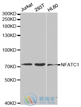
| Caculated MW: | 78 kDa;101 kDa |
| Observed MW: | Refer to Figures |
| Applications: |
WB 1:500-1:2000 IHC 1:50-1:200 IF 1:50-1:200 |
| Reacitivity: | Human, Mouse, Rat |
For research use only. Not intended for diagnostic or therapeutic use!
Additional information